
Mitosis and Meiosis⁚ A Study Guide
Mitosis and meiosis are two of the most commonly misunderstood topics on the AP Biology exam. This complete review guide will give you a crash course in mitosis and meiosis stages, and highlight the key differences between mitosis and meiosis. It will also teach you how to study through suggested… Mitosis and meiosis⁚ comparative characteristics. The fate of chromosomes during mitosis and meiosis. The fate of the cytoskeleton during mitosis and meiosis. This set of clear, simple DIAGRAMS and STUDY GUIDE will help your Biology students visualize chromosome movements during MITOSIS and MEIOSIS. And, the concise 2-page study guide summarizes the key events of the processes.
Introduction
Cell division is a fundamental process in all living organisms, ensuring growth, repair, and reproduction. There are two main types of cell division⁚ mitosis and meiosis. Mitosis is responsible for the growth and repair of somatic cells (body cells), while meiosis produces gametes (sex cells) for sexual reproduction. This study guide will delve into the intricacies of both processes, outlining the key differences between mitosis and meiosis, and highlighting the importance of each in the context of life.
Key Differences between Mitosis and Meiosis
Mitosis and meiosis, while both forms of cell division, differ significantly in their purpose and outcomes. Mitosis produces two identical daughter cells, each with the same number of chromosomes as the parent cell. This process is essential for growth, development, and repair. Meiosis, on the other hand, produces four daughter cells, each with half the number of chromosomes as the parent cell. This reduction in chromosome number is crucial for sexual reproduction, ensuring that offspring inherit a balanced set of genetic material from both parents.
Mitosis⁚ The Process of Cell Division
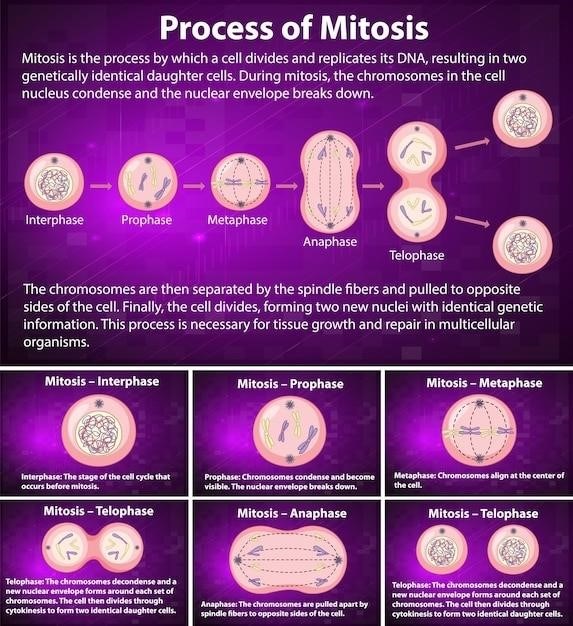
Mitosis is a fundamental process in all living organisms, responsible for the growth, development, and repair of tissues. It is a type of cell division that produces two identical daughter cells from a single parent cell. This process involves the duplication of the parent cell’s chromosomes, followed by their separation into two distinct nuclei. Mitosis ensures that each new cell receives a complete and accurate set of genetic material, maintaining the genetic integrity of the organism.
Prophase
Prophase is the first and longest stage of mitosis, characterized by the condensation of chromatin into visible chromosomes. During this stage, the nuclear envelope breaks down, allowing the spindle fibers to attach to the chromosomes at their centromeres. The spindle fibers, made of microtubules, are responsible for separating the chromosomes during the later stages of mitosis. Prophase is a crucial stage for ensuring the proper alignment and separation of chromosomes, ensuring that each daughter cell receives a complete set of genetic material.
Metaphase
Metaphase, the second stage of mitosis, is characterized by the alignment of chromosomes at the center of the cell, forming the metaphase plate. The spindle fibers, attached to the centromeres of each chromosome, pull the chromosomes towards opposite poles of the cell, ensuring that each daughter cell receives a complete set of genetic material. This precise alignment is crucial for the equal distribution of chromosomes during cell division, preventing genetic errors and maintaining the integrity of the genome.
Anaphase
Anaphase, the fourth stage of mitosis, is characterized by the separation of sister chromatids, the two identical copies of a replicated chromosome. The centromeres of each chromosome split, and the spindle fibers shorten, pulling the sister chromatids apart towards opposite poles of the cell. This process ensures that each daughter cell receives one copy of each chromosome, maintaining the correct number of chromosomes for proper cell function. The separation of sister chromatids during anaphase is a critical step in ensuring the accurate distribution of genetic material to the daughter cells.
Telophase
Telophase is the final stage of mitosis, marking the completion of nuclear division. During this stage, the chromosomes, which have been separated and pulled to opposite poles of the cell, begin to uncoil and relax, returning to their less condensed chromatin form. The nuclear envelope, which had broken down during prophase, reforms around each set of chromosomes, creating two distinct nuclei. The mitotic spindle fibers disassemble, and the cytoplasm divides, a process known as cytokinesis, which ultimately results in the formation of two daughter cells, each with its own nucleus and a complete set of chromosomes. Telophase ensures that each daughter cell receives a complete set of genetic information, essential for the proper functioning of the newly formed cells.
Meiosis⁚ The Process of Gamete Formation
Meiosis is a specialized type of cell division that occurs in sexually reproducing organisms, specifically in the germ cells, which are the cells that give rise to gametes, such as sperm and egg cells. The primary function of meiosis is to reduce the chromosome number by half, ensuring that the offspring inherit one set of chromosomes from each parent. This process involves two consecutive divisions, Meiosis I and Meiosis II, resulting in the production of four haploid daughter cells, each containing half the number of chromosomes found in the original diploid cell. Meiosis is crucial for maintaining genetic diversity, as it allows for the shuffling and recombination of parental chromosomes, contributing to the unique genetic makeup of each offspring.
Meiosis I
Meiosis I is the first of two meiotic divisions, and it’s characterized by the separation of homologous chromosomes, resulting in two daughter cells, each containing half the number of chromosomes as the original cell. This division is further divided into four distinct stages⁚ Prophase I, Metaphase I, Anaphase I, and Telophase I. Each stage plays a crucial role in the intricate process of chromosome pairing, alignment, and separation, ultimately ensuring that each daughter cell receives a complete set of chromosomes, although with only one copy of each homologous pair.
Prophase I
Prophase I is the longest and most complex stage of meiosis I. It’s marked by the condensation of chromosomes, the pairing of homologous chromosomes (synapsis), and the exchange of genetic material between them (crossing over). This exchange of genetic material is crucial for generating genetic diversity in offspring. The paired homologous chromosomes, now called tetrads, are held together by a protein structure called the synaptonemal complex. During this stage, the nuclear envelope breaks down, and the spindle fibers begin to form, preparing for the separation of chromosomes in the subsequent stages.
Metaphase I
In metaphase I, the paired homologous chromosomes, now called tetrads, line up along the metaphase plate, which is an imaginary line that runs down the center of the cell. The spindle fibers, which are made of microtubules, attach to the centromeres of the chromosomes. Each chromosome is attached to spindle fibers from opposite poles of the cell. This ensures that when the chromosomes separate in the next stage, each daughter cell receives one chromosome from each pair. The arrangement of the tetrads at the metaphase plate is random, contributing to the genetic diversity of offspring.
Anaphase I
Anaphase I is a crucial stage in meiosis I, where the homologous chromosome pairs separate and move towards opposite poles of the cell. The spindle fibers shorten, pulling the chromosomes apart. It’s important to note that sister chromatids remain attached at their centromeres, unlike in mitosis where they separate. This separation of homologous chromosomes ensures that each daughter cell receives one chromosome from each pair, reducing the chromosome number from diploid to haploid. The random assortment of chromosomes during anaphase I further contributes to genetic diversity in the offspring.
Telophase I
Telophase I marks the completion of meiosis I, characterized by the arrival of the separated homologous chromosomes at opposite poles of the cell. The nuclear envelope reforms around each set of chromosomes, forming two distinct nuclei. The cytoplasm then divides, a process known as cytokinesis, resulting in two daughter cells. These daughter cells are now haploid, meaning they contain only one set of chromosomes, unlike the diploid parent cell. Notably, each daughter cell contains a unique combination of chromosomes due to the random assortment during anaphase I, contributing to genetic diversity.
Meiosis II
Meiosis II, the second stage of meiosis, closely resembles mitosis in its mechanics. However, unlike mitosis, meiosis II starts with haploid cells, each containing a single set of chromosomes. The key purpose of meiosis II is to separate the sister chromatids within each chromosome, creating four haploid gametes, or sex cells. Meiosis II comprises the same four stages as mitosis⁚ prophase II, metaphase II, anaphase II, and telophase II. By the end of meiosis II, four genetically distinct haploid gametes are produced, ready to participate in sexual reproduction.
Prophase II
Prophase II marks the beginning of the second meiotic division. During this phase, the nuclear envelope, which had dissolved during telophase I, reforms around the chromosomes. The chromosomes condense once again, becoming more visible under a microscope. The spindle fibers, responsible for separating the chromosomes, begin to form, extending from the centrosomes, which have duplicated during interkinesis. The centrosomes migrate to opposite poles of the cell, preparing for the separation of sister chromatids during the next stage of meiosis II.
Metaphase II
Metaphase II is characterized by the alignment of the chromosomes along the metaphase plate, which is an imaginary plane that bisects the cell. The spindle fibers, extending from the centrosomes at the poles of the cell, attach to the centromeres of the chromosomes. This attachment ensures that each sister chromatid is connected to a spindle fiber from opposite poles. This arrangement is crucial for the equal distribution of genetic material to the daughter cells during the subsequent anaphase II.
Anaphase II
Anaphase II marks the separation of sister chromatids, which were previously held together at the centromere. The spindle fibers shorten, pulling the sister chromatids apart towards opposite poles of the cell. This movement is driven by the depolymerization of microtubules, which are the building blocks of the spindle fibers. As the sister chromatids move apart, they become individual chromosomes, each carrying a complete set of genetic information. This separation ensures that each daughter cell receives a full complement of chromosomes.
Telophase II
Telophase II concludes meiosis II, marking the final stage of this division process. During this phase, the chromosomes reach the opposite poles of the cell and begin to uncoil, returning to their less condensed chromatin form. The nuclear envelope re-forms around each set of chromosomes, creating two distinct nuclei within the cell. Concurrently, cytokinesis, the division of the cytoplasm, occurs, resulting in the physical separation of the cell into four daughter cells, each containing a haploid set of chromosomes. These daughter cells are now gametes, ready for fertilization to contribute to the next generation;
Comparison of Mitosis and Meiosis
Mitosis and meiosis, while both forms of cell division, differ significantly in their outcomes and functions. Mitosis, a process of duplication, produces two identical daughter cells with the same number of chromosomes as the parent cell, ensuring growth and repair. Meiosis, however, is a reduction division, resulting in four daughter cells, each with half the number of chromosomes as the parent cell. This reduction is crucial for the formation of gametes (sperm and egg cells), which fuse during fertilization to create a new organism with a complete set of chromosomes.
Applications and Importance
The understanding of mitosis and meiosis extends beyond the realm of textbooks, finding practical applications in various fields. In medicine, understanding the intricacies of cell division is crucial for tackling diseases like cancer, where uncontrolled cell growth poses a significant threat. Researchers utilize this knowledge to develop therapies targeting the cell cycle, aiming to halt tumor progression. Furthermore, the principles of meiosis are fundamental in assisted reproductive technologies, aiding in the development of techniques like in-vitro fertilization (IVF) to overcome infertility.